YOUNGSTOWN, Ohio – Early detection is crucial in treating and surviving breast cancer. In October – Breast Cancer Awareness Month – women are encouraged more than usual to get their annual mammogram, the standard order of care to find masses in breast tissue as early as possible.
And in recent years, 3D mammography has become the “standard of care and a much more utilized tool,” says Steve Davenport, chief operating officer of Southwoods Health.
For 3D mammograms, a series of X-rays are taken of the breast tissue from all angles, which are then compiled into a single, movable image that allows the radiologist to better detect potentially cancerous tissue. By comparison, 2D mammograms consist of four X-rays: one from the top and one from the front for each breast.
Beyond just offering doctors better views from which they can glean more information, 3D mammography can also help with patients’ general wellness.
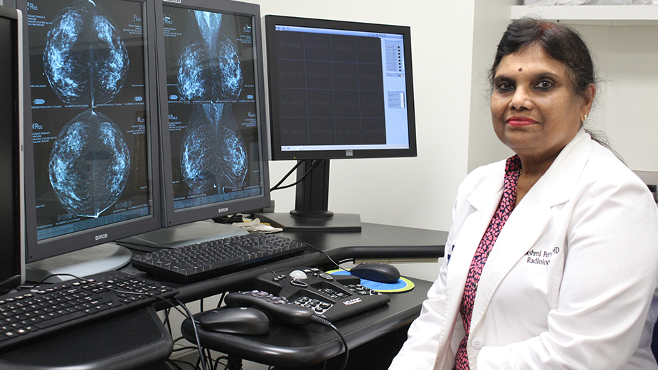
“The 3D is good because, compared to 2D, there are fewer callbacks,” whether to retake images or for more diagnostic procedures, explains Dr. S. Lakshmi Perni, a diagnostic radiologist for Steward Health Care. “When we call, part of our job is to calm them down. When they get a call back, they often think they have cancer and there’s panic. Calling back doesn’t mean you have cancer. Most of the time they’re benign.”
But while mammograms are often one of the most common tools used in screening and diagnosing breast cancer, the technology doesn’t stand alone. Radiologists can also take advantage of a swath of other tools – most commonly ultrasounds and MRIs.“Think of it like being a plumber or a carpenter: there’s all these different tools in the toolbox. In my toolbox, there are all these different ways to see the breast to identify different things,” says Dr. Alexis Smith from Mercy Health.
And like a carpenter’s tools, each imaging technology is best suited for certain tasks. Mammograms are particularly useful in finding calcifications in the tissue, while ultrasounds can help oncology teams determine if a mass is solid or cystic. MRIs, meanwhile, are “the Cadillacs of imaging,” Smith says.
“It doesn’t require radiation. If it’s negative, you know there’s nothing in the breast. That’s the problem-solving tool,” she says. “We can’t use it all the time because of the cost. But for people who are high-risk or we’re trying to solve an issue, then we’ll do an MRI.”
While the procedure can be costly, Southwoods’ Davenport notes that prices have been trending downward and that many insurance companies will cover MRIs for high risk patients. To determine this, most health care systems have patients fill out a questionnaire – covering things like age, obstetric history, family history of breast or ovarian cancer and past benign breast conditions – that assigns a score, indicating which level of care and screenings are needed.
Low-risk patients are advised to get annual mammograms starting at age 40. Intermediate-risk patients should get mammograms and, if they have dense breast tissue, ultrasounds. High-risk patients, meanwhile, are advised to get annual mammograms and MRIs, Smith explains.
“MRI provides yet another completely different image of the breast,” says Angela Kerns, Southwoods’ chief nursing officer, one that’s functionally the same as an MRI on any other part of the body. “When that risk crosses a certain threshold – usually about 15% or 20% risk – then your insurance company will likely pay for a breast MRI to stay on top of things.”
Combined, these tools allow radiologists to detect breast cancer earlier than ever. The smallest tumor that Perni has ever found, she says, was two millimeters – about half the thickness of a nickel. When first-stage breast cancer is found and treated, the survival rate is 99%, according to the American Cancer Society.
“I’ve been reading mammograms since 1984 and catching them small means [patients’] survival,” Perni says. “When they’re that small, you can take it out and do radiation to be cured. When they’re big, that’s when mastectomies and chemotherapy come in.”
Beyond just detection, the imaging tools can also help guide treatment. Quality mammograms, ultrasounds and MRIs can help surgeons get better biopsies, which in turn can produce more accurate results and avoid the need for surgery to investigate a mass.
“In the past, you might not be able to see the lesion. You knew it was there, but you couldn’t find the core, which would return an inconclusive result. Then, that would lead to surgical intervention so there could be a definitive diagnosis,” Kerns says. “By getting that first biopsy right, we can save someone from having to get that incision.”
Images taken during screenings and diagnoses are also used to help plan treatments, including surgical interventions, chemotherapy and radiation therapies.
“It’s a very big deal to have accurate imaging that shows everyone involved in the full spectrum of what’s going on. You have to know everything in order to treat it right,” Smith says.
“We look at the images together and decide on a treatment plan by taking everything into account, from pathology to what was seen under a microscope to what was seen in the imaging to what the surgeon saw during a physical exam. All of that has to be taken into account when you’re treating cancer.”
Pictured: Among the tools radiologists like Dr. S. Lakshmi Perni use to find breast cancers are mammograms, ultrasounds and MRIs.